A monoclonal cell line is essentially generated from an original polyclonal population, but it is required to separate from the polyclonal pool in order to create a pure, monoclonal cell line that is genetically identical. This is a cost-effective yet tedious process, however, a homogeneous monoclonal cell line can be obtained in theory and stable suspension cell strains can also be screened.
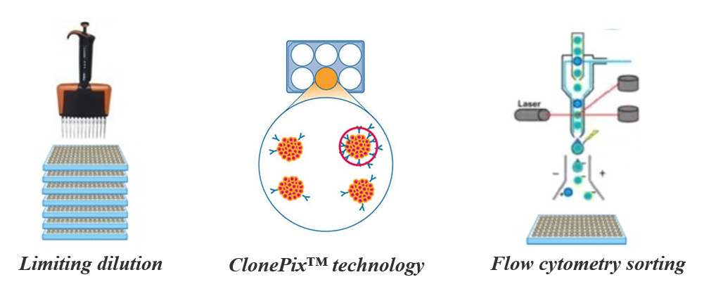
There are some technologies for generating monoclonal cell lines such as limiting dilution, ClonePix™ technology and flow cytometry sorting. The following technical procedures describe two methods that were used to successfully generate single cell clones. (The protocol is modified from the article
The art of generating single cell clones, Chelsey Kline, 2019)
Limiting dilution method
The goal of this method is to isolate each individual cell that carries selection by plating them at very low cell densities (< 1 well per well in 96 well plates) and expand colonies from those single cells in separate wells. The experiment procedure of the limiting dilution method is as follows:
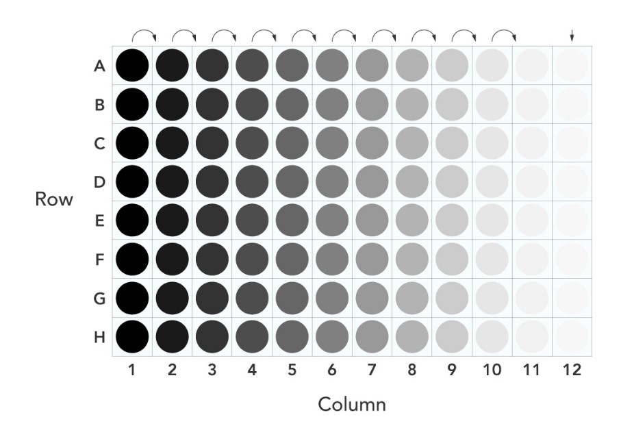 Figure 1. Plate setup for serial dilution method. Note that a 2-fold serial dilution is performed horizontally across the plate.
1. Plate ~16,000 cells polyclonal cells from the selection step in a 96-well tissue culture plate adding 200 µl per well,the other 95 wells should contain 100 µL of the respective medium. Perform the procedure at least 2 96-well plates in order to generate at least 10-20 clones.
2. Remove 100 µL from A1 containing cells and mix with B1, pipetting up and down three times to mix well before removing 100 µL and mixing with C1, etc. until reaching H1, which now has 200 µL of medium.
3. Quickly add 100 µL of medium to A1-G1 (so that all wells in column 1 will contain a total of 200 µL). Using a multichannel pipette, dilute the cells 1:1 across the rows starting with column 1, mix up and down three times, and then move to column 2 and so forth.
4. After the dilutions, wells are filled with another 100 µL of medium before being incubated. All wells now have 200 µL of medium.
5. This approximates that at least 15 wells should contain 1 cell. However, this is not always perfect and therefore monitoring the wells are very important to ensuring 1 cell per well.
Note:The dilution step can be repeated a second time. Repeating the limited dilution step 3-4 times ensures that there are no false-positive monoclones. After the monoclonal lines have been sufficiently expanded, screen the lines for transgene expression and/or other phenotypes. For example, perform Western blotting to screen for lines with the highest or lowest transgene expression.
Cloning rings/cylinders:
In this method, selected cells are seeded sparsely but not at limiting dilution in 10 cm dishes and allowed to expand and form discernible colonies for 2-3 weeks. The individual colonies can then be trypsinized and transferred to another smaller culture vessel using either cloning rings or trypsin discs for monoclonal expansion.
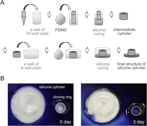 Figure 2. Preparation of the attachable silicone cylinder. (Park HB, Biochem Biophys Res Commun. 2016)
1. Count the cells so that there are no more than 20 cells per 10cm dish. This allows the cells to be spread out in the dish.
2. The next day, mark the single cells that have attached to the plate and seem far enough away from the other cells. Remember: the cells will need to be monitored for clonal expansion.
3. Once there are a few single cell colonies in the dish, use the cloning ring to isolate them.
4. To do this, carefully aspirate the medium from the dish. Using forceps, take the sterilized cloning ring that has grease on one side and place it very carefully around the tiny growing colony of single cell clones.
5. Add approximately 40 µL of trypsin into the ring and incubate the cells for about five minutes in order to carefully detach the cells. Wash and resuspend the cells into a new dish with spent medium to allow them to grow and expand.
Note: The method of cloning ring does not have a barrier and therefore cannot prevent cells in one colony from migrating and growing with another colony; this may result in not having a true single cell clone. Thus monitoring the cells to generate a single cell clone is so important.
If you have any questions,please email us at sales@brainvta.com.
 sales@brainvta.com