Fluorescent proteins are often used in our experiments, but how to select the appropriate one? Hopefully this post will give you some recommendation in which fluorescent proteins to choose.
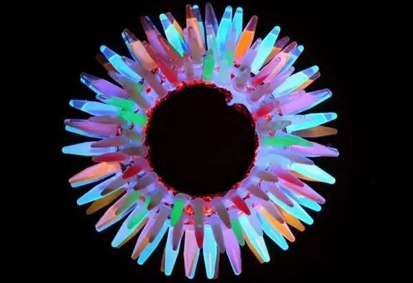
 Tubes of various fluorescent proteins displayed in a box with UV light shining on them.
1. Excitation/Emission wavelength
Each fluorescent protein has its own unique excitation/ emission peaks. Therefore, choose the fluorescent proteins with the appropriate excitation/emission wavelength that your system can excite. When using more than one fluorescent proteins, you need to choose the fluorescent proteins that can be distinguished. An accurate determination of whether two fluorescent proteins can be separated from each other requires know their excitation and emission spectra, and a good rule of thumb is that both the peak excitation wavelengths and peak emission wavelength of the two proteins should be separated by 50-60 nm.
Table1.The excitation/emission wavelength of the most popular fluorescent protein variants.
Protein
(Acronym) |
Excitation
Maximum
(nm) |
Emission
Maximum
(nm) |
|
GFP (wt) |
395/475 |
509 |
|
Green Fluorescent Proteins |
|
EGFP |
484 |
507 |
|
Emerald |
487 |
509 |
|
Superfolder GFP |
485 |
510 |
|
Azami Green |
492 |
505 |
|
TagGFP |
482 |
505 |
|
TurboGFP |
482 |
502 |
|
AcGFP |
480 |
505 |
|
ZsGreen |
493 |
505 |
|
Blue Fluorescent Proteins |
|
EBFP |
383 |
445 |
|
EBFP2 |
383 |
448 |
|
mTagBFP |
399 |
456 |
|
Cyan Fluorescent Proteins |
|
ECFP |
439 |
476 |
|
mECFP |
433 |
475 |
|
mTurquoise |
434 |
474 |
|
CyPet |
435 |
477 |
|
AmCyan1 |
458 |
489 |
|
TagCFP |
458 |
480 |
|
mTFP1 (Teal) |
462 |
492 |
|
Yellow Fluorescent Proteins |
|
EYFP |
514 |
527 |
|
Venus |
515 |
528 |
|
mCitrine |
516 |
529 |
|
TagYFP |
508 |
524 |
|
Orange Fluorescent Proteins |
|
Kusabira Orange |
548 |
559 |
|
Kusabira Orange2 |
551 |
565 |
|
mOrange |
548 |
562 |
|
mOrange2 |
549 |
565 |
|
dTomato |
554 |
581 |
|
dTomato-Tandem |
554 |
581 |
|
TagRFP |
555 |
584 |
|
TagRFP-T |
555 |
584 |
|
DsRed |
558 |
583 |
|
DsRed2 |
563 |
582 |
|
Red Fluorescent Proteins |
|
mRuby |
558 |
605 |
|
mApple |
568 |
592 |
|
AsRed2 |
576 |
592 |
|
mRFP1 |
584 |
607 |
|
JRed |
584 |
610 |
|
mCherry |
587 |
610 |
2. Oligomerization
Some fluorescent proteins are prone to oligomerization. When expressed in fusion with the target gene, it may affect the biological function of the target gene protein. Therefore, it is recommended to use monomeric fluorescent proteins(usually denoted by a “m” as the first letter in the protein name, such as mCherry) when tracking or locating target proteins.
3. Oxygen
The maturation of the chromophore on many fluorescent proteins (particularly those derived from GFP) requires oxygen. Therefore, these fluorescent proteins can only be used in oxygen sufficient environment. Now, of course, scientists have developed fluorescent proteins that can mature in an oxygen-deficient environment. Therefore, the researcher needs to consider the environment when choosing a fluorescent protein.
4. Temperature
The maturation times and intensity of fluorescent proteins can be affected by temperature. For instance, enhanced GFP (EGFP) is most suited for mammalian or bacteria studies, as it is optimized at 37 °C, whereas GFPS65T is better suited for yeast studies (24-30 °C).
5. Photostability
Fluorescent molecules will be bleached (i.e. lose the ability to emit light) after prolonged exposure to excitation light. In ordinary fluorescence imaging, the photostability of fluorescent proteins is not required to be very high. For example, in confocal laser imaging, the stability of the fluorescence signal only needs to be able to ensure a complete image collected, while for the superresolved fluorescence imaging, it is required a high photostability of the fluorescent proteins.
6. pH Stability
This parameter is important when you are planning to choice the fluorescent proteins. Each fluorescent protein has its most suitable pH, that is, the fluorescence intensity is the highest at a certain pH. Some fluorescent proteins can glow normally under acidic conditions (e.g. mcherry) while others will be quenched (e.g. GFP).
7. Maturation Time
Fluorescent proteins need to transcription, translation, folding and other steps before they create the chromophore. The folding process takes up most of the time, which is called the maturation time. When labeling short-lived proteins, we need to consider the maturation time of the fluorescent protein. In case the target protein starts to degrade, the fluorescent protein has not yet maturation, resulting in the inability to track and locate the target protein. Therefore, the maturation time should be taken into account when selecting fluorescent proteins.
8. Optimized codon
Codons have species preference. Most of the fluorescent proteins come from Marine organisms. After codon optimization, these fluorescent proteins are more suitable for expression in mammalian cells. However, codon preference should be taken into account when need to express fluorescent proteins in other species.
9. N- or C-terminus
After selecting the appropriate fluorescent protein, we need to determine which terminal (N-terminal or C-terminal) needs to be placed, and it depends on which end of the target protein is involved in protein folding. For instance, if the C-terminal of the target protein will be folded into the inner side, the fluorescent protein needs to be placed on the N-terminal of the target protein, otherwise the fluorescence signal will not be obtained. Likewise, the fluorescent protein cannot be placed in the terminal where it will be cut. If a new protein is being studied, it is recommended to perform N-terminal and C-terminal fusion expression respectively to determine which is the best choice.
These are some of the main factors that we need to consider when choosing fluorescent proteins. If you have any questions, please email us at
sales@brainvta.com.
References
1. Zhang MS, Fu ZF, & Xu PY (2016) Extending the spatiotemporal resolution of super-resolution microscopies using photomodulatable fluorescent proteins. J Innov Opt Heal Sci9(3).
2. Zhang MS, et al.(2012) Rational design of true monomeric and bright photoactivatable fluorescent proteins. Nat Methods9(7):727-U297.
3. Costantini LM, Fossati M, Francolini M, & Snapp EL (2012) Assessing the Tendency of Fluorescent Proteins to Oligomerize Under Physiologic Conditions. Traffic13(5):643-649.
4. Zhang X, et al.(2016) Highly photostable, reversibly photoswitchable fluorescent protein with high contrast ratio for live-cell superresolution microscopy. P Natl Acad Sci USA113(37):10364-10369.
5. Li D, et al.(2015) Extended-resolution structured illumination imaging of endocytic and cytoskeletal dynamics. Science349(6251).
6. Wang S, et al.(2016) GMars-Q Enables Long-Term Live-Cell Parallelized Reversible Saturable Optical Fluorescence Transitions Nanoscopy. Acs Nano10(10):9136-9144.
7. Kilaru S, et al.(2015) A codon-optimized green fluorescent protein for live cell imaging in Zymoseptoria tritici. Fungal Genet Biol79:125-131.
